









Malignant tumors are diseases with high clinical mortality rates, prompting scientists to continuously search for more effective drugs to control or even cure cancer. For a long time, drug screening for tumors relied on traditional two-dimensional (2D) cultures and patient-derived xenograft (PDX) models. However, in drug screening, 2D cell lines lack optimal physiological relevance, and immortalized cell lines grown in vitro or transplanted in vivo have been shown to exhibit minimal similarity to their original tissues or tumors. Although PDX models, derived from patients, retain the structure, cellular composition, and molecular characteristics of the original tumors, their high cost and lengthy timelines limit their application in precision cancer therapy. Therefore, scientists in the field of cancer research have long been committed to developing better three-dimensional (3D) research models.
The advent of 3D cell culture technology has provided researchers with cell models that more closely mimic the real in vivo environment. Its advantages include better simulation of the cellular microenvironment, cell-cell interactions, and biochemical and physiological responses. Leveraging these strengths, 3D cell culture technology has demonstrated outstanding performance in drug development, stem cell culture, organ regeneration, and other research fields. In 2009, Hans Clevers and colleagues made a groundbreaking advancement in intestinal organoid culture systems, ushering in a "new era" of organoid technology. Scientists discovered that primary tumor tissues could also be used to construct tumor organoids. Compared to simple 3D spheroids, the multicellular nature of organoids makes them more patient-relevant and capable of predicting patient responses to drugs. Moreover, organoid models are compatible with standard in vitro assays, such as cell viability testing.
Explore the Future of Cancer Research with the KeyTec® Luminescent 3D Cell Viability Detection Kit.
Fast and Easy
High Sensitivity and Stability
Make Your Scientific Research More Precise!
KeyTec® Luminescent 3D Cell Viability Detection Kit is based on the ATP bioluminescence method and employs efficient homogeneous lysis technology to accurately quantify the number of viable cells in 3D cultures. Compared to traditional 2D detection reagents, its enhanced lysis system thoroughly penetrates microtissue barriers, perfectly addressing the permeation challenges of complex models such as organoids and 3D spheroids, ensuring accurate and reliable data. While retaining the conventional ATP detection principle, this kit is specifically optimized for the lysis performance of 3D cultures. It is easy to operate and compatible with high-throughput detection, making it an ideal tool for tumor research, drug screening, and related fields.
* Strong Lysis Ability
The kit significantly improves lysis capability, with powerful permeability for microtissues, making it highly suitable for cell viability detection in 3D spheroids and organoids.
* Simple and Fast
Ready to use—just open and add reagents. Data can be read within 30 minutes or less, enabling highly efficient 3D cell viability detection.
* High Sensitivity
This kit is optimized from the standard KeyTec® Luminescent Cell Viability Detection Kit, perfectly retaining the sensitivity of the 2D cell viability detection kit.
* Long Half-Life
The luminescent signal generated by the kit is highly stable, with a half-life typically reaching 3 hours, making it suitable for high-throughput screening assays.
* Excellent Storage Stability
The kit undergoes rigorous testing. After thawing, the reagents can be stored at 2–8°C for 60 days (with≥90% activity). Truly ready to use—no need for repeated freezing.
The kit uses D-luciferin and thermostable mutant luciferase in the lysis solution to lyse cell clusters and react with intracellular ATP. Luminescence readings can quantitatively measure viable cells within a certain cell count range.

Figure 1. KeyTec® Luminescent 3D Cell Viability Detection Kit principle
Data Presentation
.png)
.png)
Figure 2. The KeyTec® Luminescent 3D Cell Viability Detection Kit detects the cell viability of tumor organoids (e.g., lung cancer, thyroid cancer) with readings outperforming imported brands.
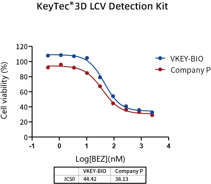
Figure 3. The KeyTec® Luminescent 3D Cell Viability Detection Kit measures drug activity in tumor organoids, with IC50 values comparable to competitors.

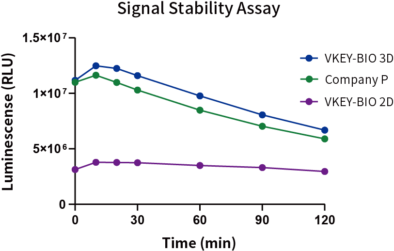
Figure 4. The KeyTec® Luminescent 3D Cell Viability Detection Kit detects Hela cell spheroid models: high signal values, a half-life exceeding 2 hours, meeting high-throughput demands.
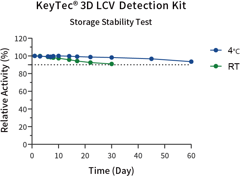
Figure 5. The KeyTec® Luminescent 3D Cell Viability Detection Kit achieves stable storage at 4°C for at least 60 days with >90% performance retention.
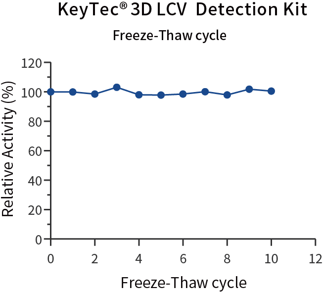
Figure 6. The KeyTec® Luminescent 3D Cell Viability Detection Kit shows no significant performance impact after 10 freeze-thaw cycles.
KeyTec® 3D Cell Viability Detection Kit features strong lysis ability, simple operation, and excellent stability, making it highly suitable for cell viability detection in 3D spheroids, organoids, and other microtissues. If you are looking for a reliable and efficient 3D cell viability detection tool, welcome to apply for a trial! VKEY-BIO is ready to collaborate with you to drive scientific breakthroughs and advance progress in the life sciences.
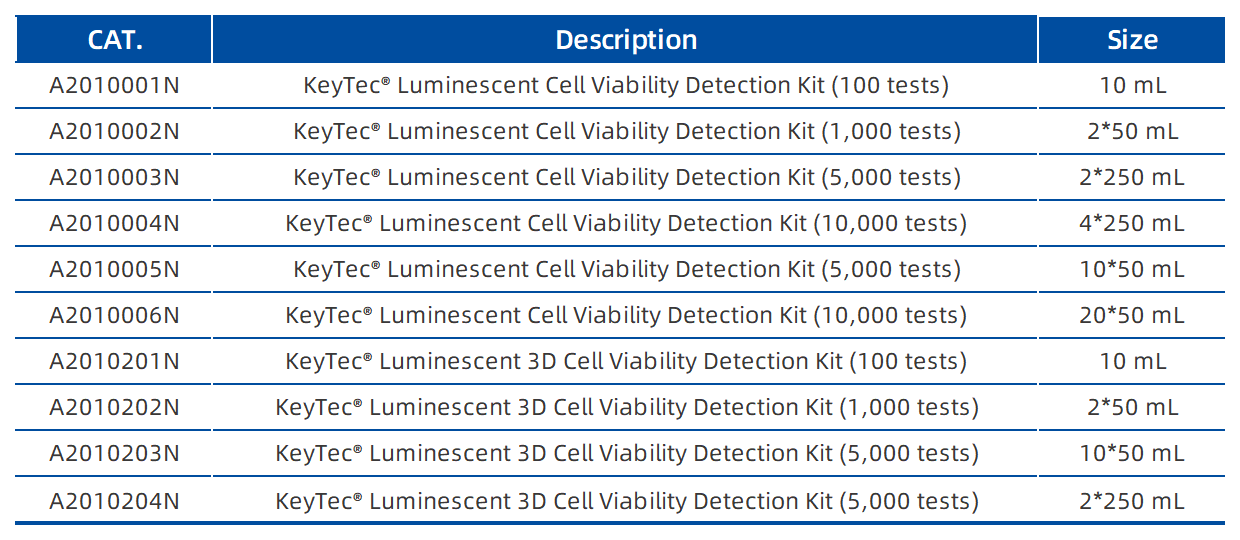
VKEY-BIO Cell Viability Detection Kit Product List:
